Black Mold vs Green Mold vs Pink Slime - What the Color Tells You
Black Mold vs. Green Mold vs. Pink Slime: What the Color Tells You
Reading time: ~6 minutes | Mold Diagnostics Color Identification Health & Safety
Why Color Matters Diagnostically
Microorganisms that colonize wet plastic or glass produce visible pigments for a range of biological reasons — UV protection, predator deterrence, metabolic byproducts. The pigment is a side effect of biology, not an aesthetic choice, which is why it's a reasonably reliable identifier of which organism is producing it. The colour, combined with the texture (fuzzy, slimy, chalky) and the location inside the jug, narrows the possibilities considerably.
This article walks through the colours most commonly seen in residential and office water jugs — what each one likely is, what it isn't, and what the right cleaning response is for each. Use this as a starting reference, not a final diagnosis. If you have a serious health concern about exposure, a laboratory swab and ID is the only fully accurate identification.
Black Spots: Often (But Not Always) Mold
Black spots inside a water jug are most commonly the genus Cladosporium, one of the most widespread household molds. Cladosporium is dark green to nearly black depending on lighting, with a slightly fuzzy texture under magnification. It thrives at room temperature with steady moisture — exactly the environment a water jug provides. Less commonly, black spots can be Aspergillus niger (which is more of a sooty black) or, in rarer cases, Stachybotrys chartarum (the well-known "black mold" — which is uncommon in water containers because it prefers cellulose-rich substrates like drywall).
The popular media has made "black mold" synonymous with Stachybotrys and an exaggerated health threat. The reality is that black mold in a water jug is almost always Cladosporium, which is a known allergen but not in the same toxicity category as Stachybotrys. The health implications of mold in drinking water depend on the species and individual sensitivity.
What black spots aren't: black spots can also be mineral deposits (iron-bearing scale produces dark staining in some hard-water households), or extremely advanced biofilm (mature biofilm darkens as bacterial populations diversify). Both look like mold from a distance but feel different on close inspection.
Green or Olive Spots: Cladosporium or Penicillium
Green spots — ranging from olive to grey-green to bright blue-green — are typically Cladosporium in earlier growth phases, or Penicillium, the genus that gave the world penicillin. Penicillium in particular is often a vivid blue-green, with a powdery surface texture, similar to what you'd see on aging citrus fruit.
Both of these are extremely common indoor molds that spread through airborne spores. Their presence in a water jug usually traces to spore introduction during refill or jug change, with germination occurring in the warm, moist interior over days or weeks. Neither is acutely dangerous to most adults at the contamination levels typical of home water jugs, but both can trigger allergic reactions in sensitized individuals.
Pink-to-Orange Slime: Usually Not Mold at All
Serratia marcescens produces a pinkish-red pigment called prodigiosin, which gives the bacterial colony its distinctive colour. It's environmental rather than usually pathogenic, but it's an indicator of biofilm establishment and warrants the same level of cleaning intervention as mold. Some people are sensitive to Serratia exposure — particularly those with respiratory or urinary conditions — so the appropriate response isn't "it's just bacteria, ignore it." It's "treat the contamination, but understand what you're treating."
White Fuzzy Spots: Mold (Usually Penicillium or Aspergillus)
White or off-white fuzzy patches are typically early-stage Penicillium or Aspergillus colonies, before they've developed their mature pigment. These appear as small cottony tufts on the interior surface, often near the waterline. White mold is the easiest to underestimate because it doesn't have the visual alarm of black spots — but it represents the same kind of fungal contamination and needs the same treatment.
White fuzzy spots are not the same as white residue. White mineral residue is flat, chalky, and crystalline. White mold has texture and dimension; you can see it has structure under good light.
Yellow or Yellow-Green Stains: Bacteria or Aged Biofilm
Yellow staining can come from several sources: pigmented bacterial colonies (some Pseudomonas species produce yellow pigments), iron-related staining in well-water households, or mature biofilm that has progressed to a yellow-brown phase. Less commonly, yellow can be early Aspergillus growth.
If yellow staining is broadly distributed across the jug rather than concentrated in spots, biofilm or mineral staining is more likely than mold. If it's localized to specific spots with raised texture, fungal growth is more likely.
The Color-to-Treatment Map
| Visible Color | Most Likely Cause | Treatment Approach |
|---|---|---|
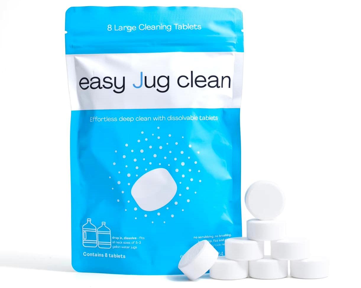
| Black, fuzzy spots | Cladosporium (mold) | 2 Easy Jug Clean tablets, 30 min, repeat next day |
| Green, fuzzy spots | Cladosporium / Penicillium (mold) | Same as black mold protocol |
| White, fuzzy patches | Early Penicillium / Aspergillus (mold) | Same as established mold protocol |
| Pink/orange slime | Serratia marcescens (bacteria) | 2 tablets, 20 min, single treatment usually sufficient |
| Yellow staining (broad) | Bacterial biofilm or mineral staining | 2 tablets, 20 min; chelating agents handle scale |
| Yellow spots (localized, fuzzy) | Aspergillus (mold) | 2 tablets, 30 min, repeat next day |
| Black flat staining (no texture) | Iron mineral deposits or mature biofilm | 2 tablets, 20 min; chelation removes iron staining |
| White flat residue (chalky) | Mineral scale (calcium/magnesium) | 1 tablet, 20 min; chelating agents dissolve it |
Why the Same Treatment Works for All These Organisms
Despite the diagnostic differences, the cleaning chemistry that handles each of these contamination types is fundamentally the same: active oxygen oxidation paired with chelation and surfactant action. Mold cells, bacterial cells, biofilm matrices, and mineral scale all respond to this combination because the active ingredients target structures common to all of them — biological membranes, polysaccharide scaffolds, and ionic mineral bonds.
When to Get Professional Identification
For most residential cases of visible contamination, the colour-based first diagnosis above plus a thorough Easy Jug Clean treatment is sufficient. Three situations warrant professional swab testing or environmental consultation:
- Recurring contamination after multiple proper treatments. If mold-appearing spots return within weeks despite consistent cleaning, the species or source pathway may need professional identification.
- Health symptoms in household members that may relate to mold exposure — chronic respiratory issues, unexplained allergies, GI symptoms in immunocompromised individuals.
- Office or commercial settings where multiple users are affected. Workplace water cooler contamination can become a public health matter requiring documented identification.
✅ One Protocol Handles Black, Green, Pink, White, and Yellow
The colour identifies the contamination. The cleaning chemistry handles all of them. Two tablets in warm water, 20–30 minutes, rinse. No species-specific products required.
FAQ: Mold and Slime Color Identification
Q: I'm seeing multiple colours in the same jug. Does that mean multiple organisms?
Often yes. Mature contamination in a long-neglected jug typically hosts multiple species — bacterial biofilm with mold colonies growing on top, sometimes with mineral staining in the background. The treatment is the same; the message is just that the contamination has been established for some time.
Q: My family member said the dark spots look red, not black. Could that change the diagnosis?
Possibly. Red-purple staining can be Serratia in heavier concentration, certain iron-related deposits, or some specific bacterial species. Lighting affects perception of colour in a contained jug interior. Treat as you would for visible contamination of any colour.
Q: After treatment, the spots are gone but there's faint discoloration left. Is that still mold?
Often residual staining in the plastic itself — pigment that the mold or bacteria deposited during growth. Active oxygen removes the organisms; some pigment may remain on the substrate. Not active contamination, but a sign the contamination was significant. Inspect for any active growth (texture, fuzziness, smell) over the following weeks.
The Bottom Line
Colour is a diagnostic clue, not a verdict. Black, green, white, pink, yellow — each suggests a likely organism category, and each warrants the same active oxygen treatment. The pillar guide walks through full mold removal; this article helps you read what you're looking at before you treat.


 Order now — get it by